
Elite Hair Centers
We know that choosing a hair restoration provider is an important decision. At Elite Hair Centers, our unique dermatology and plastic surgery team is dedicated to delivering exceptional results with expertise and passion. This is what sets us apart from the competition.
Tinea Capitis
Tinea capitis is a scalp fungal infection that leads to scaling and hair loss. It is mainly caused by dermatophyte fungi, which spread through direct contact with infected humans, animals (e.g., cats, dogs, cows, guinea pigs), or contaminated objects (e.g., combs, brushes, hats). Some infected carriers may not show symptoms. The fungi infect the hair follicle, resulting in three infection patterns: endothrix, ectothrix, and favus. In endothrix infections, the fungal spores are found inside the hair shaft. In ectrothrix infections, the fungal spores are found outside the hair shaft. And in favus, hyphae and air spaces are found within the hair shaft.
In the United States, Trichophyton tonsurans is the most common causative agent, followed by Microsporum canis. T. tonsurans spreads from person to person, while Microsporum is transmitted from animals to humans.
Tinea capitis typically affects prepubescent children and may resolve spontaneously at puberty. However, it can also occur in adults over 60. The reasons for this are not fully understood but could be related to post-pubertal sebum production and hair follicle colonization by commensal yeast, like Pityrosporum.
Five major variants of tinea capitis exist:
- Scaly patches with alopecia – single or multiple scaly, red patches that enlarge over time with hair loss and itching.
- Patches with black dots – single or multiple patches with black dots representing broken hairs at the scalp level.
- Widespread scaling with subtle hair loss – diffuse scaling without significant hair loss, resembling seborrheic dermatitis or psoriasis.
- Kerion – severe, pus-filled folliculitis with multiple inflammatory pustules, thick crusting, a boggy mass, and often associated with lymphadenopathy.
- Favus – a unique presentation with a yellow, cup-shaped crust developing around the hair follicle, potentially leading to permanent scarring and hair loss.
In addition to examination of the scalp, diagnosis may involve checking for palpable cervical lymphadenopathy, using dermoscopy, potassium hydroxide (KOH) preparation, fungal culture, polymerase chain reaction (PCR) testing, or a scalp biopsy.
Treatment typically requires systemic antifungal medications, as topical agents have limited penetration into the hair follicle. Oral griseofulvin has been the first-line treatment for children, but terbinafine is an alternative. Fluconazole and itraconazole are less commonly used but can be effective. When an infection is confirmed, all family members should be evaluated with scalp cultures to reduce the risk of recurrence.
Adjunctive therapies, such as 2% ketoconazole shampoo, 2.5% selenium sulfide shampoo, and 1% ciclopirox shampoo, can be used 2-3 times per week to prevent fungal shedding. Close contacts should follow similar preventative shampooing regimens. Thoroughly disinfect shared brushes, combs, and bed linens with bleach. If treated appropriately, hair usually regrows fully.

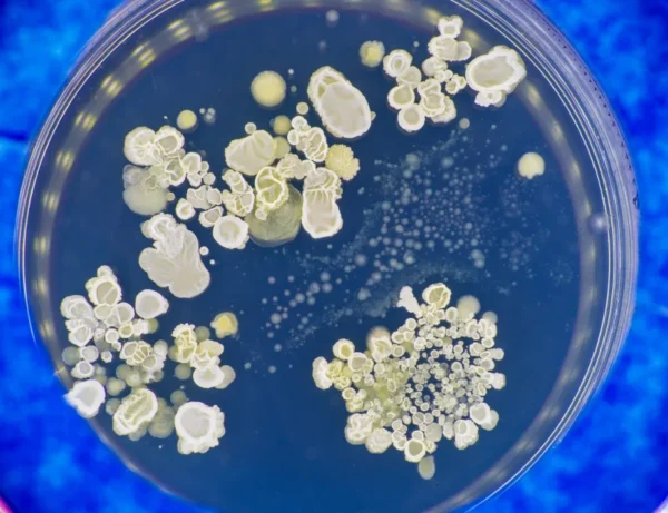
KOH Scraping
PHOTOS
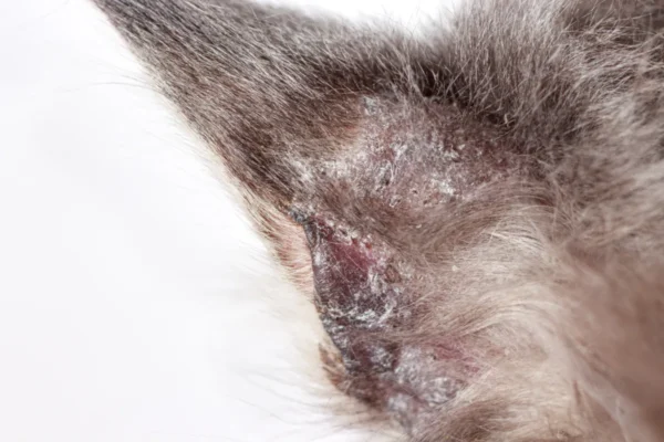
Infected cats can be a source of infection.
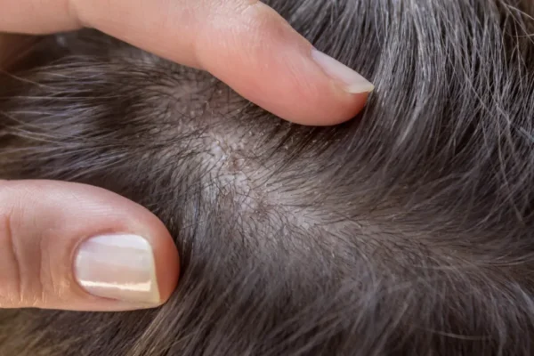
Typical appearance on the scalp.
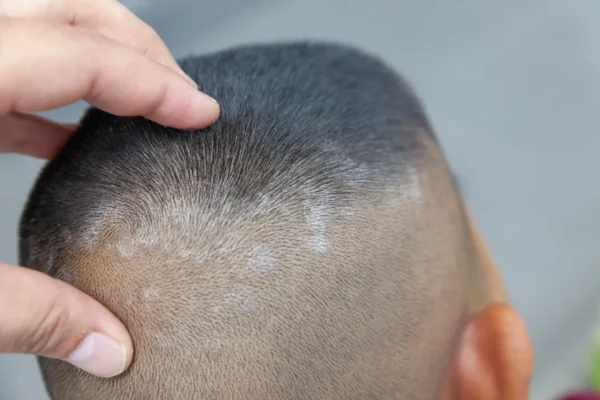
Typical appearance on the scalp.
TESTIMONIALS

Before & After Gallery
*Individual results may vary.