
Elite Hair Centers
We know that choosing a hair restoration provider is an important decision. At Elite Hair Centers, our unique dermatology and plastic surgery team is dedicated to delivering exceptional results with expertise and passion. This is what sets us apart from the competition.
Alopecia Areata
Alopecia areata is a chronic, immune-mediated form of non-scarring hair loss with an unpredictable course that may sometimes prove resistant to treatment. It is believed to result from T-lymphocytes attacking one’s own hair follicles. This condition can present in various ways, from patchy hair loss in oval or circular patterns to the complete absence of scalp and body hair. In cases of patchy alopecia areata, hair regrowth usually occurs within a year without treatment. Unfortunately, treatments for alopecia areata are generally palliative rather than curative.
The lifetime risk of developing alopecia areata is around 2%, with the mean age of diagnosis in one’s mid-30s, although it can occur at any age and affects men and women roughly equally. Genetic predisposition and environmental triggers likely contribute to this condition. Studies show that 20% of people with alopecia areata have a first-degree relative with the condition, and genome-wide studies have associated several genes, including the human leukocyte antigen (HLA) gene (HLA-DQB1*03), as a marker for susceptibility. Moreover, individuals with Turner syndrome and Down syndrome are more likely to have alopecia areata. There is speculation that a gene locus on Chromosome 21 may be partially responsible for alopecia areata given the 8.8% greater frequency of the condition in individuals with Down syndrome. Several environmental factors, including infections, medications, vaccinations, and emotional stress, have been reported as precipitating episodes.
Alopecia areata has been linked to allergic rhinitis, asthma, and atopic dermatitis. According to some research, atopy affects over 40% of people with alopecia areata compared to 20% of the general population. Another connection between alopecia areata and thyroid illness has been discovered. Moreover, autoimmune disorders like lupus, rheumatoid arthritis, myasthenia gravis, scleroderma, and lichen planus have been observed in association with alopecia areata.
Alopecia areata is considered an autoimmune process in which hair follicles in the growth phase prematurely transition to the resting phase, leading to hair shedding. The mechanism of this is not entirely understood but likely revolves around the hair follicle losing its immune privilege. The hair follicle is typically protected from the immune system; however, this protection is lost in alopecia areata, resulting in a T-cell mediated immune attack on the cells in the hair bulb, leading to follicular degeneration and hair shedding.
Clinical Features
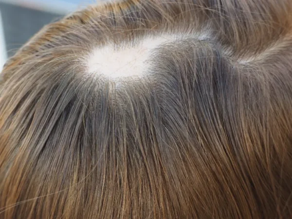
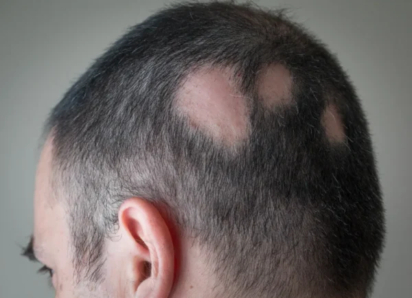
Alopecia areata most commonly produces a smooth, round or oval, completely bald patch on the scalp. Typically, there are no symptoms, although sometimes itching or burning precedes the hair loss. Both intact and broken hairs have been observed in cases of hair loss. The fragmented hairs are referred to as “exclamation point” hairs because their distal (end) section is wider than it is close to the scalp.
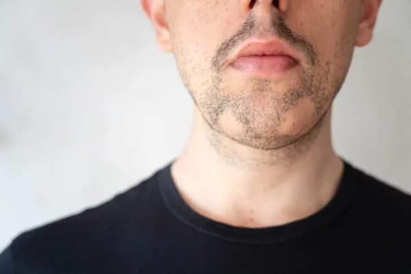
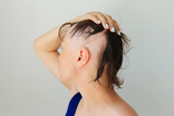
Many patients will have only discrete patches of hair loss that will completely recover within a year without therapy. However, a subset of patients (up to 10%) will progress to losing all scalp hair (alopecia totalis) or all body hair (alopecia universalis). A few unique clinical patterns exist – ophiasis, ophiasis inversus (sisapho), and diffuse. Ophiasis alopecia areata refers to a band-like area of involvement across the temporo-occipital scalp. Ophiasis inversus (sisapho) is a band-like pattern that affects the fronto-temporal scalp (opposite of ophiasis). And diffuse alopecia areata, or alopecia areata incognita, presents with abrupt, generalized thinning of the scalp hair.
Nail dystrophy may be observed in 10 to 20% of patients with alopecia areata. Dystrophy, which includes pitting and abnormal grooves (Beau’s lines), may occur before, during, or after the resolution of alopecia areata.
Approximately 50% of patients with patches of alopecia areata completely recover their hair within a year without therapy. Some patients will have persistent disease for several years or progress to more severe forms. Factors that may portend a worse outcome include cases of severe disease that start in childhood and persist for several years, a positive family history of alopecia areata, associated nail dystrophy, and the ophiasis pattern.
Histology
A biopsy of an active area of alopecia areata will demonstrate a peribulbar lymphocytic infiltrate, often referred to as a “swarm of bees.” This is a classic finding in the active periphery of alopecia areata lesions. The inflammatory infiltrate consists primarily of T-lymphocytes, along with macrophages and Langerhans cells. Additionally, an increased number of telogen hairs, miniaturization of the hairs with numerous fibrous tracts, and pigment incontinence can be observed.
TREATMENT
Currently, there is no cure for alopecia areata, and treatments aim to reduce symptoms rather than cure the disease. Local treatments may improve the appearance of the treated area but cannot prevent the spread of the condition. Because T-cell immune dysregulation is the main cause of alopecia areata, immune modulation is the primary method of treatment.
Intralesional steroid injections are the first-line therapy for adult patients with less than 50% scalp involvement. A small amount of steroid (triamcinolone acetonide 2.5 to 10 mg/cc) is injected into the dermis, and hair regrowth typically occurs 6 to 8 weeks after treatment. The treatment can be repeated every 4-6 weeks for up to 6 rounds -> and hair regrowth typically occurs 6-8 weeks after treatment. The treatment can be repeated every 4-6 weeks for up to 6 rounds, although regrowth is not guaranteed, and some patients may lose their regrowth after 12 weeks. It is important to inject the steroid intradermally or into the upper subcutis to avoid atrophy. Oral and topical minoxidil 5% can provide cosmetically acceptable hair growth in patients and is often used in combination with intralesional steroid injections. Topical immunotherapy is often a second-line treatment and involves using a contact allergen like SADBE or DPCP on the scalp to precipitate hair growth.
Systemic oral corticosteroids, cyclosporine, and methotrexate have all historically been used to treat extensive alopecia areata – all with success, but somewhat limited due to their high side-effect profiles. Recently, oral baricitinib, a selective, reversible Janus kinase 1 (JAK1) and Janus kinase 2 (JAK2) inhibitor, has demonstrated efficacy for severe alopecia areata in adults and was approved by the FDA for this purpose. Surgical hair restoration is not an appropriate treatment for alopecia areata.
TESTIMONIALS

Before & After Gallery
*Individual results may vary.